





| IN A NUTSHELL |
|
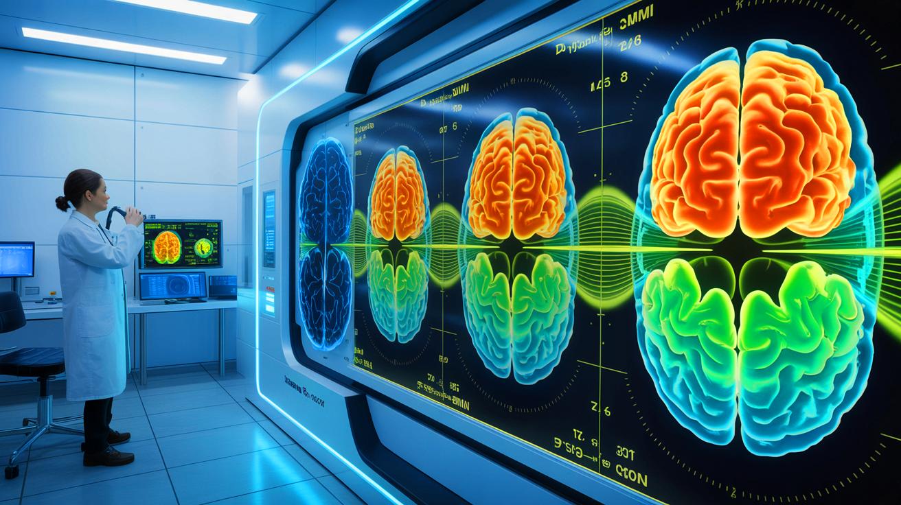
In recent years, technological advancements have revolutionized our understanding of the human brain. One of the most significant breakthroughs comes from researchers at MIT, who have developed a new imaging system that allows scientists to see deeper into brain tissue than ever before. By using light to trigger sound waves, this innovative technology can image brain tissue at depths five times greater than traditional methods. This new system, known as the multiphoton photoacoustic microscope, operates without the need for dyes or genetic modifications. This advancement opens up new possibilities for neuroscience research and surgical applications, potentially transforming how we study and treat brain diseases.
Seeing Deeper Into the Brain
The development of this advanced imaging system marks a significant leap forward in neuroscience. Traditional light-based microscopy systems have been limited in their ability to penetrate deep brain regions while maintaining resolution. The MIT system, however, can image molecules like NAD(P)H, which are critical for understanding cell metabolism and neuronal activity, through dense brain samples. These tests included a 1.1-millimeter human cerebral organoid and a 0.7-millimeter slice of mouse brain tissue.
According to W. David Lee, the postdoctoral researcher involved in the system’s design, the device employs a three-photon excitation method. This method fires ultrashort light bursts at wavelengths three times longer than the molecule’s normal absorption wavelength, reducing scattering and enabling deeper tissue penetration. Most absorbed energy results in microscopic thermal expansion, generating sound waves detectable by a sensitive ultrasound microphone. This process, known as three-photon photoacoustic imaging, converts sound waves into sharp images.
Merging Advanced Imaging Techniques
The MIT team has integrated several advanced imaging techniques into a cohesive platform termed “Multiphoton-In and Acoustic-Out.” This combination allows for precise molecular detection without altering the tissue structure. The system can identify various molecules, such as GCaMP, used for tracking neural activity. Additionally, third-harmonic generation imaging provides detailed cellular structure mapping, offering both structural and molecular insights in a single scan.
Co-lead author Tatsuya Osaki emphasized that the goal was to fuse these sophisticated techniques into one efficient process. This capability is crucial for studying conditions where NAD(P)H levels fluctuate, such as Alzheimer’s disease, Rett syndrome, and seizures. Furthermore, its label-free operation can potentially guide neurosurgical procedures by mapping brain activity in real-time, providing an invaluable tool for surgeons.
Potential Applications in Medicine and Research
While the initial results are promising, the next phase involves testing the system in living animals. This will require both the light source and microphone to be positioned on the same side of the tissue. According to Lee, the system should theoretically image up to 2 millimeters deep in live brains, a significant depth for real-time brain mapping.
Previous work by Lee through Precision Healing Inc. demonstrated the potential of NAD(P)H imaging in guiding wound treatment. This same technology could now extend its benefits to neurosurgery and brain research. The project has received support from numerous organizations, including the National Institutes of Health and The Picower Institute, highlighting its significance and potential impact.
Future Directions and Challenges
The development and successful deployment of this imaging system represent a monumental achievement in brain research and medical technology. However, challenges remain in adapting the technology for use in live subjects and ensuring its practical application in clinical settings. Researchers are optimistic about its future, hoping to refine the system further for broader use in neuroscience and medicine.
The study, published in the journal Light: Science and Applications, provides a foundation for future research and development. As scientists continue to explore the capabilities of this technology, the question remains: how will these advancements reshape our understanding of the brain and influence future medical treatments?


Incredible breakthrough! How soon can we expect this tech in clinical settings?
Wow, no dyes or genetic modifications needed? That’s a game-changer! 😊
This sounds amazing, but how cost-effective is it for widespread use?
Finally, a way to see deeper into the brain without harmful dyes. Kudos to MIT!
Does this mean we can better understand diseases like Alzheimer’s now?
Every time I read about MIT’s innovations, I feel like the future is now. 🚀
So, can this tech be used during brain surgeries in real time?
Sounds too good to be true. What’s the catch with this new system?
Is this system compatible with existing medical imaging equipment?